Project Ideas

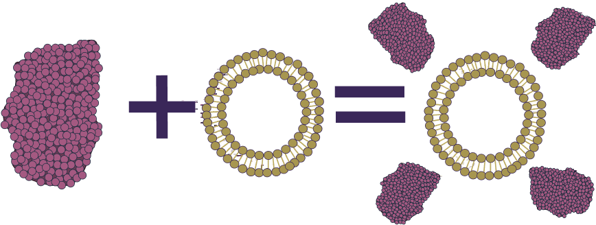
This project focuses on addressing the challenges posed by bacterial biofilms, structures that play a significant role in infectious diseases and are highly resistant to conventional antibiotic treatments. Follow along to see what factors played a role in our decisions.

Relevance

Bacterial Biofilms

A bacterial biofilm is a structure of bacteria embedded within a self-produced matrix of proteins, polysaccharides, and eDNA that often play roles in the pathogenicity of infectious diseases due to increased resistance to immune responses and antimicrobial therapy (Vetsby et al., 2020). The presence of bacterial biofilms has been associated with several severe health complications for humans. For example, cystic fibrosis is a genetic condition in which mucus builds up in the body and can induce the growth of pathogenic bacterial biofilms. Bacterial biofilms of Pseudomonas aeruginosa (P. aeruginosa) and Staphylococcus aureus (S. aureus) are two common bacterial biofilms that have also been isolated in patients diagnosed with cystic fibrosis and are credited for damaging lung tissue by causing chronic inflammation (Høiby et al., 2010) and pulmonary infections (Esposito et al., 2019). Bacterial biofilms, particularly of various Staphylococci, have also been frequently discovered on medical devices and in hospital environments, and can be detrimentally transferred to patients as they are resistant to both chemical and physical antibacterial sterilization measures, highlighting the need for more efficient and effective therapeutic treatments for bacterial biofilm infections (Zheng et al., 2018).

Conventional Therapies

Antibiotic treatments for bacterial biofilm infections in humans involve introducing drugs that will inhibit the life processes of bacteria. Currently, biofilm infections in patients are primarily treated by antibiotics either inhaled, ingested, or injected into the body via the bloodstream (Manos, 2021). However, there are several concerns with current practices including:

  • Inability of antibiotics to penetrate biofilms and organ tissues, such as requiring specific particle or droplet sizes to reach certain airways of the lungs (Høiby, 2011)
  • Historically, biofilms such as S. aureus and P. aeruginosa infections often occur concurrently, so patients receive multiple antibiotic treatments simultaneously that may adversely react and potentially pose toxicity hazards in the bloodstream (Esposito et al., 2019)
  • Concerns about biofilm antibiotic tolerance and mutational resistance undermining the ability of antibiotic treatments to eradicate biofilms on their own

Physical therapy to remove bacterial colonies in patients by expelling mucus from airways, which is commonly infected by biofilms, is often used conjointly with antibiotics. Treatments include postural drainage, percussion therapy, forced expiratory methods, and breathing techniques (Manos, 2021). Limitations include:

  • Inconclusive findings of the effectiveness of physical therapy, particularly if not performed correctly (Manos, 2021)
  • Not recommended for use on children, and some methods are physically intensive (Manos, 2021)
  • Does not directly address bacterial infections so are not suitable as the sole treatment (Manos, 2021)

Scientific Interest

To circumnavigate the challenges of antibiotics penetrating biofilms, some emerging research focuses on the prospective use of materials, such as enzymes, capable of disrupting biofilm matrices in conjunction with antibiotics (Yin et al., 2022). These enzymes work by disrupting biofilm matrices to reduce the resistance of bacteria, allowing antibiotics to take effect. Our project attempts to test the efficacy of the enzyme DNase I as part of a liposome platform to degrade biofilms in the hopes of developing a possible practical treatment for bacterial biofilm infections. DNase I is an endonuclease that is currently being studied to disrupt P. aeruginosa biofilms due to its ability to hydrolyze phosphodiester linkages present in eDNA in biofilms (Yin et al., 2022). A critical component of our project involves testing the ability and efficiency of DNase I to degrade DNA after it has been treated to conjugate with liposomes, thereby demonstrating the proof of concept and feasibility of enzymosomes for use in treatment.

Technological Interest

This project endeavors to create a method that consistently produces a delivery device capable of transporting controlled payloads of potentially useful drugs and enzymes for biofilm degradation for the treatment of bacterial infections. The ability of liposomes to be conjugated to enzymes to create a delivery vehicle capable of destabilizing biofilms was investigated. Synergistic properties of several combinations of enzymes with different functions and properties were researched to actualize a future extension of assembling a modular platform of liposomes conjugated to several enzymes capable of treating biofilm infection in a more comprehensive manner. The production method of bipyramidal DNA structures (known as “n-gonal bipyramids”) made by DNA Origami technology, and its usage as a precise template that could catalyze the formation of uniform-sized liposomes at the nanoscale was also evaluated.

Aims

To address the current limitations of treatments against antibiotic-resistant bacteria, our project aims to create conjugated enzymosome systems of DNase I enzymes and liposomes that are:

  • Able to reach regions not typically accessible to previous treatments
  • Capable of targeting and degrading biofilms
  • More uniform in size for precise and consistent drug dosages when created by a DNA n-gonal bipyramid template in the future

An overview of our enzymosome formation, in which the pink structure represents enzymes and the lipid ring represents liposomes.

Future Extensions

In the future, after we have seen positive results of the viability of our experiments with DNase I, a possible extension of our project is to test our liposome with different combinations of enzymes to design a modular enzymosome platform that could simultaneously disrupt various biofilm components. Current treatments involving DNase I face challenges of eDNA in mature biofilms resisting DNase I, possibly by binding to polysaccharides and proteins that protect from enzymatic degradation or due to reduced dependency on eDNA in aged biofilms (Yin et al., 2022). This indicates a need for various enzyme activities that not only target eDNA but also other components of biofilm matrices (Jiang et al., 2020). A modular platform composed of various enzyme constituents could allow for a more efficient destabilization of biofilms by combining the activity of different enzymes to target not only eDNA but also other key components of the extracellular matrix, such as polysaccharides and proteins. We would hypothesize that the efficacy of our enzymosomes would compound, enhancing their capability to disrupt biofilms. Two other enzymes we have researched that have been shown in past studies to have synergistic effects, and thus could be viable to work together as a unit with DNase I, are Alginate Lyase and Alpha Amylase.

  • Alginate Lyase is an enzyme that degrades alginate by cleaving the β-1,4 glycosidic bond (Zhu & Yin, 2015). In mucoid strains of P. aeruginosa biofilms, alginate has been found to be a major polysaccharide overproduced in biofilm matrices (Colvin et al., 2011). Thus, alginate lyase could be useful in reducing eDNA resistance to DNase I and assist in dispersing biofilms by weakening bonded polysaccharides. Several studies have already demonstrated a significant decrease in the survival of P. aeruginosa colonies treated with alginate lyase (Cotton et al., 2009).
  • Alpha Amylase is an enzyme capable of cleaving α-1,4-glycosidic bonds of various starches (Lahiri et al., 2021). This property may address the need to target polysaccharides bound to eDNA to increase the effectiveness of DNase I treatment in mature biofilms (Lahiri et al., 2021). Alpha amylase has also been shown to be effective at inhibiting biofilms in isolation (Craigen et al., 2011)

Presently alginate lyase has been tested in combination with both DNase I and alpha amylase. Antimicrobial treatments co-administered with both DNase I and alginate lyase have been proven to induce improved activity in reducing biofilm growth (Alipour et al., 2009). Previous experiments employing alginate lyase and alpha amylase in enzyme cocktails have indicated positive efficacy when the enzymes are used together (Kaur et al., 2021). While alpha amylase treatments in conjunction with DNase I had not been tested, a report did find that alpha amylase enzymes treated with DNase I prior to being exposed to biofilms retained their activity to destabilize biofilms (while several other enzyme treatments degraded the alpha amylase) (Kalpana et al., 2012). So, while it is unclear if they will increase the activity of one another, it is known that enzyme activity will not be impaired. Further investigation on DNase I and alpha amylase combinations would be necessary to quantify if the tandem usage yields more effective biofilm degradation as part of the enzymosome co-delivery platform.

Merit

What Sets Our Project Apart from Others?

This project is a novel combination of past research into using DNA nanotemplates to create uniform liposomes (Yang et al., 2016) and attaching enzymes to the outside of liposomes for effective delivery (Corvo et al., 2015) in order to degrade biofilms. This can be applied to a wide variety of situations where biofilms are involved, such as medical conditions like cystic fibrosis. The advantages of our project are as follows:

  • Uniform Liposomes with Controlled Enzyme Dosage

Using the DNA ring nanotemplate, we are able to create controlled uniform sizes of liposomes (Yang et al., 2016). Each liposome is saturated with a controlled dose of enzymes that will degrade biofilms, allowing for effective and predictable use. This is important because it allows for more consistency and predictability when the enzymosomes are used to treat biofilms, with a given number of liposomes always degrading the same amount of biofilm. For medical usage, such as for patients diagnosed with cystic fibrosis, this allows treatment to be accurately tailored to the amount of biofilm found in patients to completely degrade them, eliminating uncertainty about whether the biofilms have been degraded.

  • Liposomal Delivery to Regions not Reachable by Cleaning Agents

Biofilms pose a recurring problem in the area of health and medicine. From the formation of biofilms on medical devices, notably by Staphylococci bacteria (Zheng et al., 2018), to biofilm growth in the lungs of patients diagnosed with Cystic Fibrosis, they possess a diverse array of targets (Høiby et al., 2010). However, these areas may not be easily reachable, making drug delivery difficult. With the aid of a smaller form of transportation such as a liposome saturated with enzymes, these regions can become accessible to drug delivery.

Specifications

What are we Creating?

Our project aims to create an enzymosome structure that can serve as a platform for attaching enzymes and cargo. This platform is intended to enhance the efficiency of biofilm degradation in patient-related scenarios, such as cystic fibrosis, as well as in the context of environmental biofilms found on medical equipment.

The following diagram outlines the project’s proposed wet-lab timeline, with key milestones and goals marked for each month.

April

  • Research and protocol development for liposome formation, enzyme activity and enzyme conjugation
  • Beginning of de novo structure design of Trigonal and Pentagonal Bipyrimidal DNA Origami shapes
  • Replication of the design of the scaffold and staple strands of the Octahedron structure in Cadnano

May

  • Simulation of the formation stability of Octahedron using CanDO
  • Finalization of the enzyme conjugation protocols and ordering of required reagents
  • Carrying out enzyme docking simulations to pick an enzyme to test conjugation with liposome
  • Implementation of Trigonal and Pentagonal Bipyramid into Cadnano

June

  • Ordering of p7308 scaffold and staple strand sequences for octahedron structure to test in lab
  • Performing activity assays on chosen enzyme (DNAseI) in different buffer components required for conjugation (via thiolation)

July

  • Testing of different methods to produce liposomes within 100 d.nm
  • Optimize production of octahedron by testing different ratios of scaffold: staples
  • Finalize Cadnano designs of Trigonal and Pentagonal structures

August

  • Produce liposomes with and without maleimide linkages
  • Test methods of thiolating DNase I in lab performing PicoGreen activity assays to verify enzyme function
  • Run CanDo simulations on trigonal and pentagonal bipyramid structures

September

  • Attempt to prepare Octahedron samples for visualization through atomic force microscopy (AFM)
  • Purify thiolated DNase I and attempt conjugation with maleimide-liposomes
  • If resources availible, form liposome using octahedron structure

October

  • Troubleshooting for enzyme thiolation and/or conjugation
  • Finalization of data analysis of results and completion of writeups for competiion

While our project's overarching goals are ambitious, we have streamlined our methods to establish a strong foundation and proof-of-concept for future research. In terms of feasibility, our decision to concentrate solely on the conjugation of DNase I is supported by a clear rationale for enzyme selection and allows for adequate troubleshooting time. To showcase the potential for modular utilization of our structure, we also supplement experiments that cannot be conducted in the lab with models, simulations, and well-developed protocols for future studies. Through this approach, as evidenced in our lab notebook, we effectively manage the availability of resources, including reagents and equipment, necessary for the successful execution of the proposed experiments.

Design Process and Logic

Enzyme

How was the Enzyme Selected?

P. aeruginosa and S. aureus biofilms are two bacterial pathogens commonly associated with antimicrobial resistance. Antibiotic treatment often cannot eradicate these biofilm infections due to antibiotic tolerance and mutational resistance. These bacteria have the ability to form biofilms in many environments, because they are able to grow in moist conditions with simple nutritional requirements, including distilled water. Our project intended to focus on the dispersal of these two biofilms, so research into enzymes capable of degrading P. aeruginosa and S. aureus was undergone. Ultimately, DNase I was selected due to sourcing availability, substantial presence of DNA in biofilms, and synergistic effects with other enzymes that could be useful for future experiments.

EnzymeBiofilm Component TargetedOptimal pHSize
DNase I (specifically bovine pancreatic DNaseI)Hydrolyzes both single-and double-stranded DNA in extracellular matrices and on the surface of bacterial cells (Yin et al., 2022). In biofilms, eDNA is critical for early adhesion and formation of biofilms (Nijland et al., 2010).~6.5 (Kishi et al., 2001)4.5x4x3.5nm 63nm3 (Chen, 2006)
Alginate-Lyase (specifically Alg17C)Cleaves β-1,4 glycosidic bonds in Alginate (Zhu & Yin, 2015). Alginate is an exopolysaccharide often produced in the extracellular matrix of P. aeruginosa biofilms that has structural and protective functions in the biofilm (Colvin et al., 2011).~6 (Kim et al., 2012)Various sizes (Zhu & Yin, 2015)
Alpha-Amylase (specifically pancreatic α-amylase)Degrades α-1,4-glycosidic bonds of various starches (Lahiri et al., 2021). Starches are the substrate for amylase, a polysaccharide of amylose and amylopectin, commonly found in biofilm matrices (Lahiri et al., 2021).~7(Sky-peck & Thuvasethakul, 1977)7x11.4x11.8nm 941(nm3) (Larson et al., 2011)

Enzyme Conjugation Rationale

Previous proven methods of enzyme modification– specifically for liposome conjugation–were investigated, including attaching hydrophobic moieties such as fatty acid chains to enzymes and then linking them to the liposome. Examples of these methods include using a Glycosylphosphatidylinositol (GPI) lipid anchor, attaching an acyl group to cysteine residues of the target protein (S-acylation), and modifying N-terminal glycines of the protein (N-myristoylation) (Li & Qi, 2017).

Based on previous scientific articles and studies on enzyme-liposome conjugation, we planned on modifying the enzymes through thiolation. This mechanism consists of attaching a molecule with a thiol group (S-H) to the target enzyme, then reacting the enzymes with PEGylated liposomes modified with maleimide groups to yield the desired enzymosomes (Heeremans et al, 1992). Several previous studies have successfully thiolated similar enzymes while maintaining their functionality (Corvo et al, 2015) so we hypothesized this would work with our selected enzyme, DNase I.

Fig 1. Visualization of the attachment of a thiolated target enzyme to maleimide-PEG liposomes.

Another method we previously considered was acylation, which consists of attaching an acyl group (a molecule consisting of a C=O bond) to the target enzyme. This mechanism involves attaching a fatty acyl chain to the epsilon-amino (-NH2) group of a free lysine residue on an enzyme, introducing a hydrophobic tail to the enzyme that can be inserted into the liposome and produce an enzyme-conjugated liposomes. However, previous literature showed this method may not be compatible with our chosen enzyme, DNase I. This is because DNase I contains an epsilon-amino group on the amino acid Glutamine within its active site (Kishi et al, 2001), instead of on an amino group at an N-terminus active-site that enzymes compatible with this method, like Alginate Lyase, possess (Xu et al, 2018).

Liposome

What are Liposomes?

Liposomes are artificial, spherical vesicles created from cholesterol and phospholipids (Akbarzadeh et al., 2013). Due to their biocompatible properties, hydrophobic and hydrophilic characteristics, as well as their size, there is potential to use liposomes as future drug delivery systems (Akbarzadeh et al., 2013).

Liposome Synthesis Workflow

Liposomes were synthesized via a protocol for lipid nanoparticle synthesis outlined in a 2022 paper (Wang et al., 2022). During synthesis, maleimide dissolved in a solvent was introduced to the lipid stocks to add maleimide groups to the liposome necessary for later conjugation to an enzyme. Afterwards, the liposomes were evaluated to assess the size and uniformity of the final products, which was critical to ensure accuracy in subsequent steps and potential future applications such as drug loading.

Enzymosomes

What are Enzymosomes?

A liposome conjugated to one or more enzymes produces a modular platform called an enzymosome, which is often synthesized to improve the effective therapeutic activity of enzymes for prolonged periods of time–particularly in vivo (Corvo et al., 2015). Typically, when unmodified enzymes are administered in vivo, the enzymatic activity at the target site is limited due to clearance via glomerular filtration, however, in enzymosomes this complication is addressed by attaching the enzymes to liposomes that act as carrier systems with longer circulation times (Corvo et al., 2015).

Enzymosomes Formulation Workflow

The workflow began by determining enzyme activity in various solvents of our selected enzyme, DNase I, based on a Picogreen Enzyme Activity Assay and spectrometry standard protocols (ThermoFischer Scientific, 2022). This was necessary to determine the effects the solvents used in subsequent stages had on the enzyme activity of the final produced liposome. Next, the enzymes were thiolated because of the ability of thiol groups to react with maleimide groups on the liposome to form bonds between the structures (Girão et al., 2021), before being reacted with the previously synthesized PEGylated liposomes to form the enzymosome structure.

N-Gonal Bipyramids

How was DNA Origami used?

DNA origami structures were designed for future use as templates to achieve more precise liposome formation and eventual enzyme attachment. The DNA origami template structure is an n-gonal bipyramid type endoskeleton used as a design strategy to encapsulate a lipid membrane over a DNA endoskeleton scaffold, where n-gon refers to a polygon with n number of edges(n∈ℕ), however, our project only performed experiments where n=3, 4, and 5. It should be noted that, throughout our research, the term “octahedron” is used interchangeably to refer to the n=4 square bipyramid, due to the structure formed being representative of both terms. This was hypothesized to produce liposomes with more uniformity between sample sizes and shapes and to provide high structural support necessary for prolonged surface-enzyme activity, since the liposome would be encasing a precisely designed endoskeleton. The project was inspired by a paper that explored a mechanism of natural systems, such as viruses, which encapsulate their genetic material within a lipid envelope to prevent degradation during infection, to protect the structural integrity of their DNA nanostructures in vivo (Perrault & Shih, 2014). A software called caDNAno, which runs on python and uses 2D representations to design 3D DNA origami structures, was used to design our DNA endoskeleton. Previous UBC BIOMOD teams developed python scripts that check for possible complications, including DNA trapped in local energy minima during the folding process or the relative length of staple strands affecting the annealing process (Law et al., 2023). The workflow began with determining the shape of the structure and the helices layout, followed by computational design and testing structure using online simulation tools such as mrDNA and Cando. After verifying the structures computationally, they are formed in the lab using oligo-annealing protocols and verified with a variety of imaging techniques.

References

Akbarzadeh, A., Rezaei-Sadabady, R., Davaran, S., Joo, S. W., Zarghami, N., Hanifehpour, Y., Samiei, M., Kouhi, M., & Nejati-Koshki, K. (2013). Liposome: Classification, Preparation, and Applications. Nanoscale Research Letters, 8(1). https://doi.org/10.1186/1556-276x-8-102

Alipour, M., Suntre, Z. E., & Omri, A. (2009). Importance of DNase and Alginate Lyase for Enhancing Free and Liposome Encapsulated Aminoglycoside Activity against Pseudomonas Aeruginosa. Journal of Antimicrobial Chemotherapy 64(2). https://doi.org/10.1093/jac/dkp165.

Chen, W. (2006). Structure and Function of Bovine Pancreatic Deoxyribonuclease I. Protein & Peptide Letters, 13(5). https://doi.org/10.2174/092986606776819475.

Colvin, K. M., Irie, Y., Tart, C. S., Urbano, R., Whitney, J. C., Ryder, C., Howell, P. L., Wozniak, D. J. & Parsek, M. R. (2011). The Pel and Psl Polysaccharides Provide Pseudomonas Aeruginosa Structural Redundancy Within the Biofilm Matrix. Environmental Microbiology, 14(8). https://www.ncbi.nlm.nih.gov/pmc/articles/PMC3840794/

Corvo, M. L., Marinho, H. S., Marcelino, P., Lopes, R. M., Vale, C. A., Marques, C. R., Martins, L. C., Laverman, P., Storm, G., & Martins, M. B. A. (2015). Superoxide dismutase enzymosomes: Carrier capacity optimization, in vivo behaviour and therapeutic activity. Pharmaceutical Research, 32, 91–102. https://doi.org/10.1007/s11095-014-1447-7

Cotton, L. A., Graham, R. J., & Lee, R. J. (2009). The Role of Alginate in P. Aeruginosa PAO1 Biofilm Structural Resistance to Gentamicin and Ciprofloxacin. Journal of Experimental Microbiology and Immunology, 13, 58–62.

Craigen, B., Dashiff, A., & Kadouri, D. E. (2011). The Use of Commercially Available Alpha-Amylase Compounds to Inhibit and Remove Staphylococcus Aureus Biofilms. The Open Microbiology Journal, 5, 21–31. https://doi.org/10.2174/1874285801105010021

Esposito, S., Pennoni, G., Mencarini, V., Palladino, N., Peccini, L., & Principi, N. (2019). Antimicrobial Treatment of Staphylococcus aureus in Patients With Cystic Fibrosis. Frontiers in Pharmacology, 10, 849. https://doi.org/10.3389/fphar.2019.00849

Girão, L. F. C., Carvalheiro, M. C., Ferreira-Silva, M., da Rocha, S. L. G., Perales, J., Martins, M. B. F., Ferrara, M. A., Bon, E. P. S., & Corvo, M. L. (2021). ASP-Enzymosomes with Saccharomyces cerevisiae Asparaginase II Expressed in Pichia pastoris: Formulation Design and In Vitro Studies of a Potential Antileukemic Drug. International Journal of Molecular Sciences, 22(20), 11120. https://doi.org/10.3390/ijms222011120

Heeremans, J. L. M., Kraaijenga, J. J., Los, P., Kluft, C., & Crommelin, D. J. A. (1992). Development of a procedure for coupling the homing device glu-plasminogen to liposomes. BBA-General Subjects, 1117(3), 258-264. https://doi.org/10.1016/0304-4165(92)90022-M.

Høiby, N. (2011). Recent advances in the treatment of Pseudomonas Aeruginosa Infections in Cystic Fibrosis. BMC Med, 9(32). https://doi.org/10.1186/1741-7015-9-32

Høiby, N., Ciofu, O., & Bjarnsholt, T. (2010). Pseudomonas aeruginosa biofilms in cystic fibrosis. Future Microbiology, 5(11), 1663–1674. https://doi.org/10.2217/fmb.10.125

Jiang, Y., Geng, M., & Bai, L. (2020). Targeting Biofilms Therapy: Current Research Strategies and Development Hurdles. Microorganisms, 8(8), 1222. https://doi.org/10.3390/microorganisms8081222

Kalpana, B. J., Aarthy, S., & Pandian, S. K. (2012). Antibiofilm Activity of α-Amylase from Bacillus Subtilis S8-18 Against Biofilm Forming Human Bacterial Pathogens. Applied Biochemistry and Biotechnology, 167,1778–94. https://doi.org/10.1007/s12010-011-9526-2.

Kaur, A., Soni, S. K., Vij, S., & Rishi, P. (2021). Cocktail of Carbohydrases from Aspergillus Niger: An Economical and Eco-Friendly Option for Biofilm Clearance from Biopolymer Surfaces. AMB Express, 11. https://doi.org/10.1186/s13568-021-01183-y.

Kim, H. T., Chung, J. H., Wang, D., Lee, J., Woo, H. C., Choi, I. G., & Kim, K. H., (2012). Depolymerization of Alginate into a Monomeric Sugar Acid using Alg17C, an Exo-oligoalginate Lyase Cloned from Saccharophagus Degradans 2-40. Applied Microbiol Biotechnology, 93(5), 2233-2239. https://doi.org/10.1007/s00253-012-3882-x.

Kishi, K., Toshihiro, Y., & Takeshita, H. (2001) DNase I: structure, function, and use in medicine and forensic science. Legal Medicine, 3(2), 69-83 https://doi.org/10.1016/S1344-6223(01)00004-9

Lahiri, D., Nag, M., Banerjee, R., Mukherjee, D., Garai, S., Sarkar, T., Dey, A., Sheikh, H. I., Pathak, S. K., Edinur, H. A., Pati, S., & Ray, R. R. (2021). Amylases: Biofilm Inducer or Biofilm Inhibitor? Frontiers in Cellular and Infection Microbiology, 11. https://doi.org/10.3389/fcimb.2021.660048

Larson, S. B., Day, J. S., & McPherson, A. (2011). X-ray Crystallographic Analyses of Pig Pancreatic α-Amylase with Limit Dextrin, Oligosaccharide and α-Cyclodextrin. Biochemistry, 49(14), 3101-3115. https://doi.org/10.1021/bi902183w.

Law, M., Susham, C., Mackay, D., Nguyen, S., Nicholas, R., Tsai, M. R. G., Rajkumar, E., Inaba, F., Maheden, K., Abdi, I., Ho, J. C. H., Kieft, B., & Hallam, S. J. (2023). Self-Assembly of a Repeatable DNA Nanohinge System Supporting Higher Order Structure Formation. bioRxiv. https://doi.org/10.1101/2023.05.26.542516

Li, Y. & Qi, B. (2017). Progress toward Understanding Protein S-acylation: Prospective in Plants. Front. Plant Sci, 8, 346. https://doi.org/10.3389/fpls.2017.00346

Manos, J. (2021). Current and Emerging Therapies to Combat Cystic Fibrosis Lung Infections. Microorganisms, 9(9), 1874. https://doi.org/10.3390/microorganisms9091874

Nijland, R., Hall, M. J., & Burgess, J. G. (2010). Dispersal of biofilms by secreted, matrix degrading, bacterial DNase. PLoS ONE, 5(12). https://doi.org/10.1371/journal.pone.0015668

Perrault, S. D., & Shih, W. M. (2014). Virus-Inspired Membrane Encapsulation of DNA Nanostructures To Achieve In Vivo Stability. ACS Nano, 8(5), 5132–5140. https://doi.org/10.1021/nn5011914.

Sky-Peck, H.H., & Thuvasethakul, P. (1977). Human pancreatic alpha-amylase. II. Effects of pH, substrate and ions on the activity of the enzyme. Annals of Clinical & Laboratory Science, 7(4), 310-317.

ThermoFischer Scientific. (2022). Quant-iTTM PicoGreenTM dsDNA Reagent and Kit. https://www.thermofisher.com/document-connect/document-connect.html?url=https://assets.thermofisher.com/TFS-Assets%2FLSG%2Fmanuals%2Fmp07581.pdf

Vestby, L. K., Grønseth, T., Simm, R., & Nesse, L. L. (2020). Bacterial Biofilm and its Role in the Pathogenesis of Disease. Antibiotics (Basel), 9(2), 59. https://doi.org/10.3390/antibiotics9020059.

Wang, X., Liu, S., Sun, Y., Yu, X., Lee, S. M., Cheng, Q., Wei, T., Gong, J., Robinson, J., Zhang, D., Lian, X., Basak, P., & Siegwart, D. J. (2022). Preparation of selective organ-targeting (SORT) lipid nanoparticles (LNPs) using multiple technical methods for tissue-specific mRNA delivery. Nature Protocols, 18, 265-291. https://doi.org/10.1038/s41596-022-00755-x

Xu, F., Wang, P., Zhang, Y., & Chen, X. (2018). Diversity of three-dimensional structures and catalytic mechanisms of alginate lyases. Applied and Environmental Microbiology, 84(3). https://doi.org/10.1128/AEM.02040-17

Yang, Y., Wang, J., Shigematsu, H., Xu, W., Shih, W. M., Rothman, J. E., & Lin, C. (2016). Self-assembly of size-controlled liposomes on DNA nanotemplates. Nature Chemistry, 8(5), 476–483. https://doi.org/10.1038/nchem.2472

Yin, R., Cheng, J., Wang, J., Li, P. & Lin, J. (2022). Treatment of Pseudomonas aeruginosa Infectious Biofilms: Challenges and Strategies. Frontiers in Microbiology, 13. https://doi.org/10.3389/fmicb.2022.955286

Zheng, Y., He, L., Asiamah, T. K., & Otto, M. (2018). Colonization of medical devices by staphylococci. Environmental Microbiology, 20(9), 3141–3153. https://doi.org/10.1111/1462-2920.14129

Zhu, B., & Yin, H. (2015). Alginate Lyase: Review of Major Sources and Classification, Properties, Structure-Function Analysis and Applications. Bioengineered, 6(3), 125–131. https://doi.org/10.1080/21655979.2015.1030543